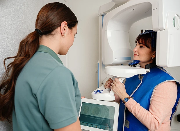
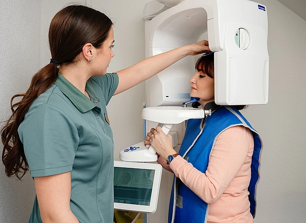
3D X-ray / digital volume tomography (DVT)

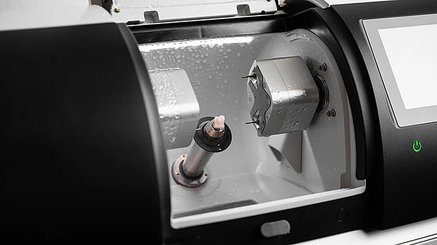
Digital volume tomography is a modern three-dimensional X-ray technique that provides highly precise images of teeth, jawbones, nerve structures, and soft tissue. Compared to conventional 2D X-rays, DVT diagnostics offer greater detail, increased safety during treatment, and significantly more precise planning, particularly in implantology, oral surgery, and endodontics.

What is 3D X-ray with DVT?

Digital volume tomography (DVT) is a modern, low-radiation, three-dimensional imaging technique that provides high-resolution, three-dimensional views of the structures in the head and jaw region.
This volumetric data allows for the precise assessment of the position, shape, density, and orientation of anatomical structures—from every angle. DVT plays an important role in:

  • Implant planning and bone analysis
  • Wisdom tooth and nerve-related surgeries
  • Root canal treatments (endodontics)
  • Cysts, fractures, and dislocations
  • Sleep apnea and temporomandibular joint diagnostics

Procedure of the DVT examination

  1. Quick preparation & positioning – the patient stands or sits in a completely relaxed position
  2. Scan process – takes only a few seconds, painless & quiet
  3. Digital analysis by the dentist
  4. Joint discussion of findings on the monitor with a 3D view

Important notes

  • The radiation exposure is lower than that of medical CT scans (computed tomography)
  • DVT is used in a targeted and indication-specific manner
  • Optimizes primarily aesthetic, surgical, and implantological outcomes
  • Supports patient-friendly decision-making through a 3D visualization of the findings